


Deepak Jangra answered this
Pituitary Gland:
Function:
- Growth Hormone Production
- Production of Hormones That Act on Other Endocrine Glands
- Production of Hormones That Act on the Muscles and the Kidneys
- Endocrine Function Regulation
- Storage of Hormones Produced by the Hypothalamus
Location:
 The pituitary gland is a small endocrine system organ that controls a multitude of important functions in the body.
- 11
m.rox98... answered this
Its present in the brain and it helps to cope up with stress.It helps in water balance andmakes endorphin to relievepain and alter mood
- 1
Shruti Anilnair answered this
pituitary gland is a pea sized gland located in the base of the skull between the optic nerves . it secretes hormones .it is referred as the master gland as it controls hormone functions such as temperature thyroid activity production of testosterone &estrogen etc.
- 3
Vedika Krishna answered this
it is at the base of brain.
it produces TSH which stimulates the growth and development of thyroid gland
It also produces GH which stimulates the growth and development of the body
- 1
Deepak Jangra answered this
Structure and Function: Pituitary Gland
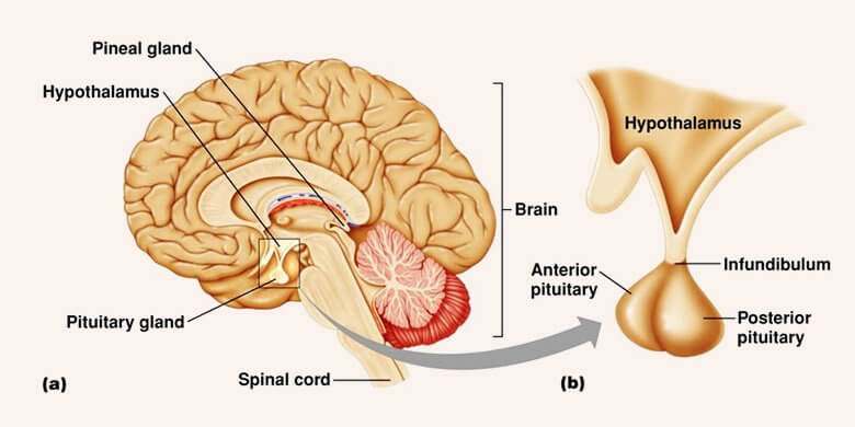
The pituitary gland controls the activities of many other endocrine glands and hormone-producing cells around the body. The pituitary gland has two parts, the anterior and posterior lobes, which are under the control of a part of the brain known as the hypothalamus. The hypothalamus secretes releasing hormones that pass through the circulation to the pituitary’s anterior lobe, where they trigger the production of hormones that control other glands. Nerve cells in the hypothalamus secrete two hormones that pass down nerve fibres to be stored in the posterior lobe until they are needed.

Structure of the pituitary gland
The pituitary gland has an anterior lobe and a posterior lobe. It is linked to the hypothalamus, which lies directly above it, by a short stalk that contains nerve fibres and a specialized network of blood vessels known as a portal system.

Pituitary hormones
Almost all of the pituitary hormones are secreted by the anterior (front) lobe of the pituitary gland, but two hormones, vasopressin and oxytocin, are produced in the posterior (rear) lobe. Pituitary gland hormones either act on other glands, stimulating them to produce hormones, or act directly on tissues and organs.
- 0
Deepak Jangra answered this
pituitary gland, also called hypophysis, ![pituitary gland: mammalian pituitary gland [Credit: Encyclopædia Britannica, Inc.]](http://media-3.web.britannica.com/eb-media/26/6526-003-38D13221.jpg) ductless gland of the endocrine system that secretes hormones directly into the bloodstream. The term hypophysis (from the Greek for “lying under”) refers to the gland’s position on the underside of the brain. The pituitary gland has a major role in the regulation of many endocrine functions.
ductless gland of the endocrine system that secretes hormones directly into the bloodstream. The term hypophysis (from the Greek for “lying under”) refers to the gland’s position on the underside of the brain. The pituitary gland has a major role in the regulation of many endocrine functions.
Table Of ContentsAnatomy of the pituitary gland
![brain: human brain, left hemisphere, medial view [Credit: Encyclopædia Britannica, Inc.]](http://media-3.web.britannica.com/eb-media/73/74273-003-6C2F8500.jpg) The pituitary gland lies at the base of the skull and is housed within a bony structure called the sella turcica. Its weight in normal adult humans is about 500 mg (0.02 ounce). The gland is attached to the hypothalamus by the pituitary stalk, which is composed of the axons of neurons and the hypophyseal-portal veins. In most species the pituitary gland is divided into three lobes: the anterior lobe, the intermediate lobe, and the posterior lobe. In humans the intermediate lobe does not exist as a distinct anatomic structure but rather remains only as cells dispersed within the anterior lobe. Despite its proximity to the anterior lobe of the pituitary, the posterior lobe of the pituitary is functionally distinct and is an integral part of a separate neural structure called the neurohypophysis.
The pituitary gland lies at the base of the skull and is housed within a bony structure called the sella turcica. Its weight in normal adult humans is about 500 mg (0.02 ounce). The gland is attached to the hypothalamus by the pituitary stalk, which is composed of the axons of neurons and the hypophyseal-portal veins. In most species the pituitary gland is divided into three lobes: the anterior lobe, the intermediate lobe, and the posterior lobe. In humans the intermediate lobe does not exist as a distinct anatomic structure but rather remains only as cells dispersed within the anterior lobe. Despite its proximity to the anterior lobe of the pituitary, the posterior lobe of the pituitary is functionally distinct and is an integral part of a separate neural structure called the neurohypophysis.
Table Of ContentsThe anterior pituitary
![pituitary gland [Credit: Uniformed Services University of the Health Sciences (USUHS)]](http://media-3.web.britannica.com/eb-media/29/106529-003-B59FD3EB.jpg) The cells constituting the anterior lobe of the pituitary gland are embryologically derived from an outpouching of the roof of the pharynx, known as Rathke’s pouch. While the cells appear to be relatively homogeneous under a light microscope, there are in fact five different types of cells, each of which secretes a different hormone or hormones. The thyrotrophs synthesize and secrete thyrotropin (thyroid-stimulating hormone; TSH); the gonadotrophs, both luteinizing hormone (LH) and follicle-stimulating hormone (FSH); the corticotrophs, adrenocorticotropic hormone (ACTH; corticotropin); the somatotrophs, growth hormone (GH; somatotropin); and the lactotrophs, prolactin.
The cells constituting the anterior lobe of the pituitary gland are embryologically derived from an outpouching of the roof of the pharynx, known as Rathke’s pouch. While the cells appear to be relatively homogeneous under a light microscope, there are in fact five different types of cells, each of which secretes a different hormone or hormones. The thyrotrophs synthesize and secrete thyrotropin (thyroid-stimulating hormone; TSH); the gonadotrophs, both luteinizing hormone (LH) and follicle-stimulating hormone (FSH); the corticotrophs, adrenocorticotropic hormone (ACTH; corticotropin); the somatotrophs, growth hormone (GH; somatotropin); and the lactotrophs, prolactin.
Somatotrophs are plentiful in the anterior pituitary gland, constituting about 40 percent of the tissue. They are located predominantly in the anterior and the lateral regions of the gland and secrete between one and two milligrams of GH each day.
Structure and function of anterior pituitary hormones
The hormones of the anterior pituitary are proteins that consist of one or two long polypeptide chains. The gonadotropins (LH and FSH) and thyrotropin are called glycoproteins because they contain complex carbohydrates known as glycosides. Each of these three hormones—LH, FSH, and thyrotropin—is composed of two glycopeptide chains, one of which, the alpha chain, is identical in all three hormones. The other chain, the beta chain, differs in structure for each hormone, thereby explaining the different actions of each of these three hormones. As is the case for all protein hormones, the hormones of the anterior pituitary are synthesized in the cytoplasm of the cells as large, inactive molecules called prohormones. These prohormones are stored in granules, within which they are cleaved into active hormones and are secreted into the circulation.
![melanocyte-stimulating hormone: pituitary gland secretions [Credit: Encyclopædia Britannica, Inc.]](http://media-2.web.britannica.com/eb-media/02/117702-003-7ADD504C.jpg) Each pituitary hormone plays a vital role in endocrine function. Thyrotropin stimulates the production of thyroid hormone. ACTH stimulates the production of cortisol and androgenic hormones by the adrenal cortex. FSH stimulates the production of estrogens and the growth of egg cells (oocytes) in the ovaries in women and sperm cells in the testes in men. LH stimulates the production of estrogens and progesterone by the ovaries in women and the production of testosterone by the testes in men. GH stimulates linear growth in children and helps to maintain bone and other tissues in adults. Prolactin stimulates milk production.
Each pituitary hormone plays a vital role in endocrine function. Thyrotropin stimulates the production of thyroid hormone. ACTH stimulates the production of cortisol and androgenic hormones by the adrenal cortex. FSH stimulates the production of estrogens and the growth of egg cells (oocytes) in the ovaries in women and sperm cells in the testes in men. LH stimulates the production of estrogens and progesterone by the ovaries in women and the production of testosterone by the testes in men. GH stimulates linear growth in children and helps to maintain bone and other tissues in adults. Prolactin stimulates milk production.
Regulation of anterior pituitary hormones
The production of the anterior pituitary hormones is regulated in part by hormones produced in the hypothalamus, the region of the brain that lies just above the pituitary gland. In general, hypothalamic hormones stimulate production of pituitary hormones, except for prolactin, which is inhibited. The hypothalamic hormones are secreted into a portal vein that traverses directly from the hypothalamus to the anterior pituitary gland, thereby carrying these hormones directly to the pituitary.
The posterior lobe is composed of the endings of nerve cells located in specialized regions of the hypothalamus. These nerve cells produce two hormones, oxytocin and vasopressin (antidiuretic hormone), that are carried down the nerves and stored in the nerve endings that compose the posterior pituitary gland. The hormones are released into the circulation in response to nerve signals that originate in the hypothalamus and are transmitted to the posterior pituitary. Oxytocin causes contraction of the uterus and milk secretion in women, and vasopressin increases reabsorption of water from the kidneys and raises blood pressure.
Table Of ContentsNeurohypophyseal system
The posterior lobe of the pituitary gland consists largely of extensions of processes (axons) from two pairs of large clusters of nerve cell bodies (nuclei) in the hypothalamus. One of these nuclei, known as the supraoptic nuclei, lies immediately above the optic tract, while the other nuclei, known as the paraventricular nuclei, lies on each side of the third ventricle of the brain. These nuclei, the axons of the cell bodies of nerves that form the nuclei, and the nerve endings in the posterior pituitary gland form the neurohypophyseal system. There are neural connections to the brain and other centres of the hypothalamus, including a centre that modulates thirst.
The two neurohypophyseal hormones, vasopressin and oxytocin, are synthesized and incorporated into neurosecretory granules in the cell bodies of the nuclei. These hormones are synthesized as part of a precursor protein that includes one of the hormones and a protein called neurophysin. After synthesis and incorporation into neurosecretory granules, the precursor protein is cleaved, forming separate hormone and neurophysin molecules, which remain loosely attached to one another. These granules are carried through the axons and are stored in the posterior lobe of the pituitary gland. Upon stimulation of the nerve cells, the granules fuse with the cell wall of the nerve endings, the hormone and neurophysin dissociate from one another, and both the hormone and the neurophysin are released into the bloodstream.
- 2
Vellai Pandi.c answered this
briefly explain the function of pituitary gland
- 2
Hartej Singh answered this
Hormones secreted from the pituitary gland help control: growth, blood pressure, certain functions of the thyroid glands and metabolism
- 1
Manasij N S answered this
- 1
Manasij N S answered this
1) Diagram of Pituitary Gland :
This answer is a brief and straight answer which I have typed on my own. Please follow this pattern of giving simple and easy answers which the asker may find easy to read and find the main content of what we try to tell him/her. And please thumbs up only if you appreciate the time i took into this and if you found it really helpful. I am sure the asker has got enough detailed info with what my other friends have given. Thank You.
- 7
Vatsalya answered this
- 0
Pushkar answered this
- 1