Can any one explain me the working,structure and functions of heartwith the help of a diagram?please

In human beings, the heart is a muscular organ. It is divided into four chambers – right auricle, right ventricle, left auricle, and left ventricle. The walls of these chambers are made up of a special muscle called myocardium, which contracts continuously and rhythmically to distribute blood to all the body cells.
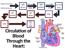
 

Working of heart

  • The heart has superior and inferior vena cava. They carry deoxygenated blood from the upper and lower regions of the body respectively and supply the deoxygenated blood to the right auricle of the heart.
  • The right ventricle contracts and passes the deoxygenated blood into the two pulmonary arteries, which pumps it to the lungs where the blood is oxygenated. From the lungs, the pulmonary veins transport the oxygenated blood to the left atrium of the heart.
  • The left atrium contracts and through the auriculo-ventricular aperture (bicuspid valve), the oxygenated blood enters the left ventricle.
  • The blood passes to aorta from the left ventricle. The aorta gives rise to many arteries that distribute the oxygenated blood to all the regions of the body. 
  • Since the blood goes twice through the heart, it is known as double circulation.


  • 25

In mammals, the function of the right side of the heart (see right heart) is to collect de-oxygenated blood, in the right atrium, from the body (via superior and inferior vena cavae) and pump it, through the tricuspid valve, via the right ventricle, into the lungs (pulmonary circulation) so that carbon dioxide can be dropped off and oxygen picked up (gas exchange). This happens through the passive process of diffusion. The left side (see left heart) collects oxygenated blood from the lungs into the left atrium. From the left atrium the blood moves to the left ventricle, through the bicuspid valve (mitral valve), which pumps it out to the body (via the aorta). On both sides, the lower ventricles are thicker and stronger than the upper atria. The muscle wall surrounding the left ventricle is thicker than the wall surrounding the right ventricle due to the higher force needed to pump the blood through the systemic circulation.

Starting in the right atrium, the blood flows through the tricuspid valve to the right ventricle. Here, it is pumped out the pulmonary semilunar valve and travels through the pulmonary artery to the lungs. From there, oxygenated blood flows back through the pulmonary vein to the left atrium. It then travels through the mitral valve to the left ventricle, from where it is pumped through the aortic semilunar valve to the aorta. The aorta forks and the blood is divided between major arteries which supply the upper and lower body. The blood travels in the arteries to the smaller arterioles and then, finally, to the tiny capillaries which feed each cell. The (relatively) deoxygenated blood then travels to the venules, which coalesce into veins, then to the inferior and superior venae cavae and finally back to the right atrium where the process began.

  • 6
What are you looking for?